

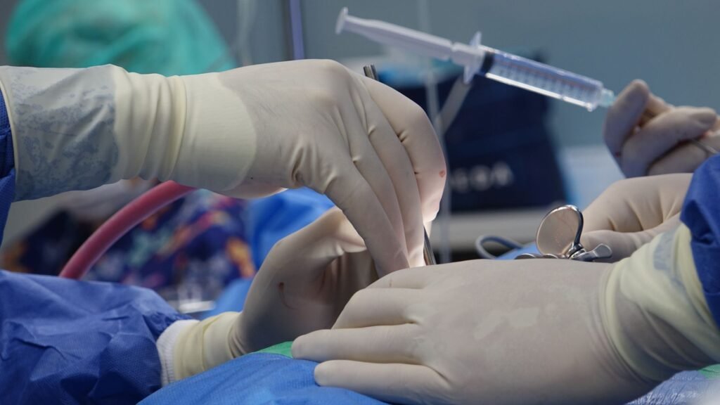
The pituitary gland is about the size of a marble. Packed around it, in a space you could cover with a postage stamp, are the carotid arteries and the optic nerves. A surgeon working there is threading an endoscope up through the nose and operating in a cavity where a millimetre in the wrong direction can mean a stroke, permanent blindness, or death.
In May, at the National Hospital for Neurology and Neurosurgery in London, a team removed a tumour from that space while an AI watched the endoscope feed and painted the dangerous structures in color as they appeared. It is being called the world’s first live AI-assisted brain surgery, and the details were made public this week. The patient, Rhys Hibbert, still has his eyesight.
The short version
- Where: the National Hospital for Neurology and Neurosurgery, part of UCLH, with research led from UCL
- Who: Professor Hani Marcus operating, with neurosurgical resident and UCL PhD candidate Danyal Khan leading the work behind the system
- The patient: Rhys Hibbert, 48, from Bedfordshire, a father of two with a non-cancerous pituitary tumour around 11 mm across
- What the AI did: analyzed the live surgical video in real time and highlighted critical anatomy at the base of the brain
- What it did not do: hold anything, move anything, or make a single decision
Why live video is the whole story
Computers have been in operating rooms for decades. Image guided neurosurgery, where a scan taken before the operation is registered to the patient’s head so instruments can be tracked against it, has been routine since the 1990s. If the headline were “computer helps neurosurgeon,” it would be about thirty years late.
The difference here is what the software was looking at. It was not consulting a scan taken last week. It was reading the same picture the surgeon was reading, frame by frame, as the operation happened.
That matters because of a problem surgeons have lived with for as long as image guidance has existed. Anatomy moves. Once you open a space, drain fluid, and start removing tissue, everything shifts. A preoperative scan is an accurate map of a place that no longer exists in quite the same shape, and the discrepancy grows the longer you work. Every experienced surgeon knows to trust their eyes over the map when the two disagree. A system that reads live video does not have that problem, because it has no map to go stale.
What “trained on more operations than a surgeon sees in a lifetime” actually means
The system was built from a large library of annotated endoscopic pituitary surgery videos, operations recorded and then labeled frame by frame so the model could learn what a carotid artery looks like from inside a nose, under a bright light, partly obscured by blood, at every awkward angle a real operation produces.
Hani Marcus described it as being trained on more operations than most surgeons will see or perform in a lifetime, and said it can act like an expert second pair of eyes. That phrasing is doing careful work, and it is worth unpacking rather than skimming.
A neurosurgeon’s expertise in this specific procedure is built from a few thousand cases at the very outside, accumulated over a career. The model has seen a superset of that, but only in one narrow sense: it has seen more frames. It has no understanding of why anything is where it is, no ability to reason about an anomaly it has never encountered, and no judgment about when to stop. What it has is pattern recognition at a volume no human career can supply, applied to one very specific visual task.
That is a genuinely useful thing to have on a screen. It is also a much narrower thing than “AI performs brain surgery,” which is roughly how half the internet has chosen to describe it.
| What people are hearing | What happened |
|---|---|
| A robot performed brain surgery | Human surgeons performed it. Nothing autonomous touched the patient |
| The AI decided where to cut | The AI colored structures on a video feed. Every decision stayed with the team |
| It used a scan of the patient’s brain | It deliberately did not. It read the live endoscope video instead |
| This is now standard NHS practice | It is government funded research with a first patient, not a rollout |
The patient, and how he got here
Rhys Hibbert is 48, works in customer services, volunteers in his community, and has two children. His tumour was found the way a lot of them are, which is to say by accident and alarmingly. He collapsed during a walk in December 2024 and had a seizure. The investigation that followed turned up a growth of roughly 11 mm on his pituitary gland.
It was not cancer. Pituitary tumours frequently are not. What makes them dangerous is location rather than biology: as one grows, it presses upward on the optic chiasm, the junction where the optic nerves cross. Left alone, the classic result is a slow loss of peripheral vision that people often do not notice until a great deal of it is gone. Without treatment, Hibbert was facing exactly that.
He seems to have found the historical symmetry more interesting than the technology. The hospital was founded in 1859 as the world’s first dedicated neurosurgical hospital, and he pointed out that it seemed fitting for the same institution to carry out the first AI-assisted neurosurgery. He described being asked to be the first patient in the research as humbling.
Where the caution belongs
One operation is one operation. The people who did it know that better than anyone, which is why this is framed as government funded research rather than a product launch. The Health Innovation Minister, James Frith, called it an example of AI at its best while adding that it needs proper safeguards, which is the sort of line ministers say but happens to be correct here.
The specific risk with a system like this is not that it fails loudly. It is that it succeeds quietly for long enough that people stop checking it. A tool that colors the carotid artery green ninety nine times out of a hundred trains the eye to expect green. The hundredth case, where the anatomy is unusual or the model has drifted outside what it was trained on, is the one where a habit becomes a hazard. Aviation spent decades learning this lesson about autopilots and gave it a name, automation complacency, and medicine gets to learn it again with different equipment.
It is also worth noting how much of the performance comes from the scaffolding around the model rather than the model itself. The same pattern showed up in a very different context recently, when a model that scored 30% on a benchmark alone hit 100% inside the right harness. Here, the harness is a surgical team, an endoscope, an annotation pipeline and a hospital’s safety culture. Take those away and there is no story.
What would make this routine
- A trial with numbers. Complication rates and extent of resection compared against matched cases without the system
- Evidence it generalizes. Different hospitals, different endoscopes, different surgical styles, different patient anatomy
- A rule for disagreement. An explicit protocol for what a surgeon does when the overlay and their own eyes do not match
The part that is genuinely new
Strip away the framing and something real remains. For the first time, a machine learning system read a live surgical field and told a surgeon what it was seeing while the surgery was still happening, in an operation where the margin for error is measured in fractions of a millimetre and the cost of an error is a person’s eyesight.
Everything downstream of that, the trials, the regulation, the arguments about which hospitals get it and when, is going to take years and should. But the technical claim underneath the headline holds up, and unlike a great deal of what gets announced in AI and medicine, this one was tested somewhere the consequences were immediate and a specific person was in the room. Brain adjacent technology has been moving quickly on several fronts at once, and we covered another one when a brain interface startup raised $21 million two months after launching. The difference is that this one already has a patient who walked out with his vision intact.
